Posts

Algae and Vibrio Fisheri

Image
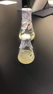
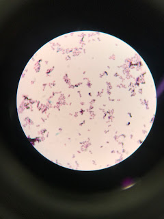
Hi! I hope everyone had a great summer! Currently, I am working on the disruption of bacterial Quorum Sensing. Quorum Sensing is how bacteria's communicate. In Vibrio Fisheri (V. Fisheri ) it controls a behavior such as bioluminescence. Also, I just started working with C. Reinhardtii (algae). Algae produce compounds that mimic N-acy homoserine lactone (AHL). V. Fisheri is grown in a PB agar plate and algae was grown in a liquid TAP . I still haven't gone to this step, but the next step will be to add algae to the center of each plate. This will cause the center of the plate to stop growing. It's the AHL blocking the QS signals who controls the bioluminescence.

Making Media + Gram stain of D. Radiodurans

Image
On 4/7, I was shown how to make media. Media is to nourish the bacteria. They live in it and eat it as well. The ingredients that were used to make media was:  Tryptone, 1.25 grams Yeast extract, 1.25 grams Dextrose (Glucose), .25g  Water, 250ml  A flask was place on a stirring hot plate and it was turn to a medium speed and 100 ml of water was added. Add the ingredients one at a time. Lastly, add the remaining water, the 150 ml, which makes it a total of 250 ml of water. I was just shown how to make media, so I didn't do anything with it once it was made.  Also,  I did a gram stain on D. Radiodurans. A gram stain is when you want to know whether a bacteria is gram-positive or gram-negative. Gram- positive bacteria have a thick layer of peptidoglycan in its cell wall, so its able to retain the crystal violet, making it look purple. After staining D. Radiodurans, it was found to be gram positive. 

Micropipettes

Image
On 3/30, I was shown how to use micropipette correctly and their correct measurements. The reason for the micropipette is to transfer small amounts of liquid (< 1ml). The scale on the micropipette are in microliters, so 1000 ul = 1 ml. Also, when using two different volumes with micropipette, the largest volume is always added first and then the smallest volume. There are four pipettes I was shown: P20, P100, P200 and P1000.  Each one has a low volume and a high volume. In the picture, the red one is the P20;  the limits are 2 ul to 20 ul. The colorless one is P100: the limits are 20 ul to 100 ul The P200, which is the yellow: the limits are 50 ul to 200 ul Lastly, the P1000 is the blue one: the limits are 200 ul and 1000ul.

Gram Stain

Image
Hi everyone! My name is Claudia Millanes. On Monday, 3/19, I perform a Gram stain of Sonorensis. I wanted to see if it was Gram-positive (GP) or Gram-negative (GN). The difference between a Gram-positive and a Gram-Negative is the structure of the cell wall. A Gram-positive baccteria has no outer cell membrane, but it's found in Gram-negative bacteria. It was concluded that Sonorensis is indeed a Gram-positive bacteria. When looking under the microscope, you can see that the bacteria retained the crystal violent dye, which was actually one of dyes being used. This is due to the high amount of peptidoglycan in the bacteria's cell wall. However, when looking at the results, there are two different types of shapes, meaning that it was contamiated.