Gram Stain
Hi everyone! My name is Claudia Millanes.
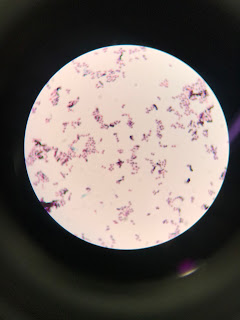
On Monday, 3/19, I perform a Gram stain of Sonorensis. I wanted to see if it was Gram-positive (GP) or Gram-negative (GN). The difference between a Gram-positive and a Gram-Negative is the structure of the cell wall. A Gram-positive baccteria has no outer cell membrane, but it's found in Gram-negative bacteria.
It was concluded that Sonorensis is indeed a Gram-positive bacteria. When looking under the microscope, you can see that the bacteria retained the crystal violent dye, which was actually one of dyes being used. This is due to the high amount of peptidoglycan in the bacteria's cell wall. However, when looking at the results, there are two different types of shapes, meaning that it was contamiated.


Great post! Good summary explaining the process of a gram stain. Excellent photo!
ReplyDeleteThat's really cool to be able to identify whether a bacteria is gram positive or gram negative. I think this information helps pharmaceutical companies develop medication for microbes using this concept. Since gram positive have a high amount of pepdidoglycan they can easily absorbs the dye, where as gram negative don't absorb as easily and have to be stained differently. Gram negative have lps on their surface, but aren't able to absorb as easy.
ReplyDeleteNice gram stain! I know they can being really hard to make when you are just starting out. I look forward to working with you in lab and learning this stuff together.
ReplyDelete